The Grams
You have seen us mention gram-positive and gram-negative everywhere now. However, what is gram-positive and gram-negative?
Basically, gram-positive bacteria have a thick peptidoglycan layer, while gram-negative bacteria have a thin peptidoglycan layer. However, gram-negative bacteria have an outer membrane.
Novato is a gram-positive bacteria.
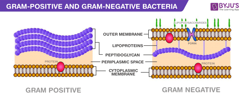
Image 1: Bacteria Cell Wall and Cell Membrane (Byjus, n.d.)
Gram Positive
Gram-positive bacteria have a thick peptidoglycan layer in front of the inner membrane. However, they DO NOT have an outer membrane.
The thick peptidoglycan layer is "super" thick, with around 40 to 80 layers of peptidoglycan molecules. Because of its chemical structure nature, peptidoglycan can easily absorb foreign substances.
However, due to the absence of a hydrophobic outer membrane, foreign substances directly contact the peptidoglycan, which absorbs them easily. This includes antibiotics, which implies that gram-positive bacteria are easier to kill!
Good news! Novato is a gram-positive bacteria.
Gram Negative
Gram-negative bacteria have a thin peptidoglycan layer in front of the inner membrane and an outer membrane in front of the peptidoglycan layer.
The thin peptidoglycan later is only 1 to 3 layers thick! However, gram-negative bacteria have an ace up their sleeve: The outer membrane. The outer membrane is composed of the phospholipid bilayer and an extra lipopolysaccharide (LPS) layer (both of which are hydrophobic).
Gram-negative bacteria are more resistant to antibiotics because of the outer membrane barrier.
LPS are extended chains of sugar molecules. They are hydrophobic, which prevents water and foreign material from penetrating the bacteria. This includes antibiotics; it is harder for antibiotics to penetrate the bacteria because of the hydrophobic area produced by the LPS (the water is not directly next to the bacteria, which is how antibiotics float to the bacteria). It is the same as Lysozyme enzymes. They cannot get near the bacteria to break the cell wall due to the hydrophobic area. Hence, the enzyme has a more challenging time reaching the bacterial surface to break the cell wall.
In addition, gram-negative bacteria have a higher concentration of transportation proteins than gram-positive bacteria. A high concentration of transportation proteins can force harmful drugs out of the bacteria before they reach core elements like the nucleus.
Gram Staining
Gram staining, a well-known lab procedure developed in the 1880s, can be used to classify gram-positive and gram-negative bacteria.
This bacteriological laboratory technique uses a crystal violet stain and checks the remaining color.
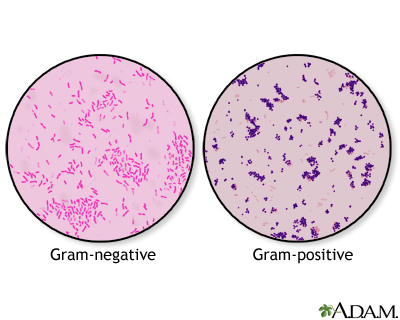
Image 2: Gram Staining Results (ADAM, n.d.)
If the remaining color is violet, it is a gram-positive bacteria, as the peptidoglycan absorbed the stain.
If the remaining color is pink, it is a gram-negative bacteria, as the thin peptidoglycan and the outer membrane cannot absorb and retain the stain.
Let us take a deep dive into the bacteriological testing procedure:
- Prepare an uncontaminated slip of bacteria sample.
- Apply a crystal violet primary stain onto the slip.
- Heat fix the bacteria slip using fire.
- This process will kill some bacteria.
- But this prevents the bacteria from washing away in the following steps.
- Add iodine on the slip.
- Rinse the slip with some ethanol drops to decolorize the bacteria.
- Finally, counterstain the slip with Safranin () and check the result.
If the slip's color is violet, then the bacteria is gram-positive. If it's pink, then it's gram-negative.